Dear colleagues,
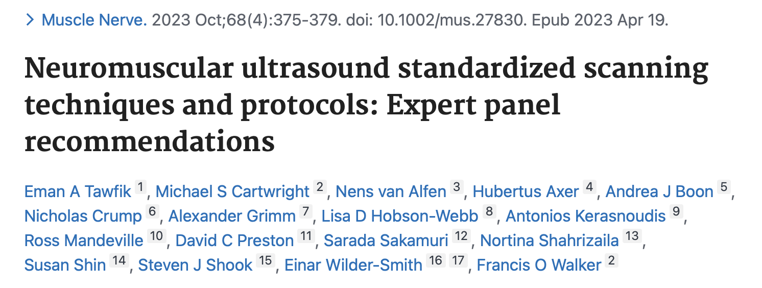
We would like to inform you about an article in the October issue of Muscle & Nerve entitled

as we thought this might be of interest for you. Furthermore, Doris Lieba-Samal had the honour of contributing to an editorial on this article and discuss the suggestions. The editorial can be provided upon personal request.
Expert panel recommendations are extremely important on our way to define how we examine, what we look for and how we document ultrasound findings to get the best results for our patients, although we do not have a solid evidence base for all of these decisions. The vivid discussion is what brings us forward in the topic we love.

The expert panel consisted of 17 internationally renowned experts on neuromuscular ultrasound. The goal of the study was development of consensus-based guidelines on:
- The ultrasound probe and type of handling
- General machine settings
- General scanning rules
- Scanning protocols for suspected focal mononeuropathy
- Scanning protocols for suspected brachial plexopathy
- Scanning protocols for suspected polyneuropathy
- Scanning protocol for suspected amyotrophic lateral sclerosis
This was achieved by a Delphi-approach with three rounds of voting on an electronic survey, that was modified after each round.

The first three protocols covered the technical details.
1a. Ultrasound probe and type of handling
Ultrasound probe and type of handling included e.g. the call for linear probes for superficial and curvilinear for deep nerves and how the probe should be held (between thumb and index and middle fingers with the ulnar side of the hand touching the patient´s skin).
1b. General machine settings
General machine settings recommend at least 12 MHz probes – which is something, we also have been promoting here. E.g. the depth is recommended so that the majority of the target structure occupies the majority of the screen and the focus should be at the level of the target nerve.
1c. General scanning rules
General scanning rules talk for example about when you should scan the entire course of the nerve (failure to localize focal lesion at the expected pathology site, suspected tandem lesion, MMN and CIDP), what should be documented in nerve ultrasound (e.g. two views – as we also recommend, CSA – if necessary comparing to the contralateral side) and in muscle ultrasound (e.g. muscle echotexture, involuntary muscle movements during rest).
2. Scanning protocols for suspected focal mononeuropathy
The scanning protocols for suspected focal mononeuropathy (entrapment and traumatic nerve injury) line out how to document them – and are in line with our recommendations of setting up a neuromuscular ultrasound lab.
3. Scanning protocols for suspected brachial plexopathy
Scanning protocol of plexopathies was rather loosely defined in our opinion. It recommends to start scanning at any preferred level and look for enlargement, which should be defined as diffuse at all levels or localized to one level.
We discussed this as probably difficult for those new in neuromuscular ultrasound. We assume levels were defined in the supraclavicular region. Then the most common differentiation is cervical/foraminal – interscalene – supraclavicular levels. However also supra- and infraclavicular levels could be addressed.
Our recommendation for starting out is in supraclavicular region, as this is the one most frequently affected in dysimmune neuropathies and trauma..
4. Scanning protocols for suspected polyneuropathy
Scanning protocol for polyneuropathies is divided into a general approach and then further steps for special situations and parameters that should be assessed (e.g. nerve echotexture, hypervascularity, external structures that could compress the nerve).
For axonal polyneuropathy the panel recommends examination of upper and lower limb nerves. This was a point of discussion in the editorial, as we would not use the dichotomy axonal – demyelinating but rather potentially treatable – not treatable, which covers dysimmune neuropathies and vasculitic neuropathy. As far as we know today, the most common polyneuropathies do not show sonographic abnormalities that go beyond the changes observed in routine electrodiagnostics.
Furthermore, scanning of the sural nerve if symptomatic is advised. This is something that also leaves room for discussion, as we know from studies that the sural nerve and the lower limb nerves in general do not aid the diagnosis of dysimmune neuropathies and the recommendation may be misleading.
5. Scanning protocols for suspected amyotrophic lateral sclerosis
Scanning protocol for suspected amyotrophic lateral sclerosis recommends scanning of the genioglossus and proximal and distal muscles of the upper and lower limb. Assessment should cover echotexture (if normal values are available), muscle thickness and fasciculations. The panel does not give any recommendation for how long or on how many sites should be scanned. We would usually do 3 sites for 30 seconds each. Then the protocol asks to determine whether there is nerve thinning – which is something that is discussed in the editorial as rather hard to achieve, as there is no consent on lower cut-off values in nerve CSA. Assessment of the brachial plexus is recommended if there is suspicion of MMN.
6. Scanning protocols for suspected myopathy
The scanning protocols for suspected myopathies recommends scanning of at least one distal and one proximal muscle in one symptomatic upper and lower limb. The assessment should include muscle echotexture and pattern of distribution, grading of muscle echogenicity (Heckmatt scale) and muscle thickness.
That´s the latest news from our side. We hope that was helpful for you. Have a good time and keep scanning!


Recent Comments