
Dear colleagues!
So you have decided to set up your own neuromuscular ultrasound lab? Congratulations on that step! We promise you will not regret it. Whether you are a physician working with electrodiagnostics already or a sonographer dealing with MSK-patients – NMUS will add a new dimension to your possibilities of non-invasive and timely diagnosing of your patients correctly.
As building an ultrasound lab from scratch is not something most of us have ever done, we decided to put this little guide together, which tells you the most important points you should think about before starting out.
As an educational platform dedicated to advancing neuromuscular ultrasound we focus on you and your needs for implementing this tool in your lab. We are here for advice and education at any stage. Just get in touch with us. Together we will get your lab started.
For the next two months we will have a weekly series running, going through all the chapters you have to consider when starting your own lab. With tips and tricks you can use right away.
This guide will also be uploaded to our Additional Resources in the next week – which means you can download and print it there for free 🙂
If you have comments or ideas – we are happy to hear them!
Now let´s get started. Fasten your seatbelts and get ready for our first chapter:

1. LOCAL AND PERSONAL SETTINGS
a. The light and the dark
Ultrasound is a dark job. When you are performing ultrasound, you are reading the images live, which means you should have optimal conditions for getting the best results quickly. Reading ultrasound images is based on differentiating grayscale levels – in other words: contrast.
Our eye has a great ability to adjust contrast to different light situations. Just think of turning off the light in an otherwise dark room. First you see nothing at all and then contours of furniture become visible again within a minute. If you then turn on the light again, you are suddenly blinded and everything is just bright, you may have to close your eyes for a moment, then adjusting again to brightness. However, the process of adapting to different light situations may take up to minutes for optimization.
It has been suggested that radiologists allow their eyes to adapt to brightness for 15 minutes before they start to work!
Your ultrasound screen has a fixed brightness, usually something about 40 lux. If your room light is much brighter (or much darker) your eye has constantly to adjust to the different settings. This makes live reading of images of course a lot more difficult and will also cause fatigue of your eyes.
It is therefore recommended, that the brightness in your ultrasound lab equals the brightness of the screen for optimal working conditions.

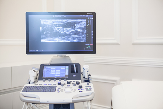
Fig. 1. Glare on the left side of the screen caused by a window

Fig. 1. Specters on the left side of the screen caused by a reading light.
Besides brightness also positioning of the light source is important. Light can get reflected on the screen, either in shape of specters or a glare, which of course again influences your ability to interpret images. If you need some light for e.g. reading prior reports, it is recommended to place the light source behind the screen to avoid disturbing reflections.
Once you got your lightning ready, let´s take the next step.
b. Postioning the patient and yourself
Most of your patients will be lying in a supine position with adjustment of their extremities according to the nerves and muscles you are interested in.
An examination couch is therefore a must. As in most cases either the left or the right side are examined, we advise you to be able to access the patient from both sides, as the examined extremity should be next to you (reaching over the patient to the other arm is not recommended). Therefore, you can mainly choose between two options:
(Note: in the following suggestions the arrows indicate the range of movement of the ultrasound machine and examiner)
The roll-over

If you have enough space, a patient centered approach may be your best choice.
The examination couch is in the center of your room, a chair with wheels on each side of it.
The advantage is that you can always position the patient in the same direction, with her or his head at the top end of the examination couch, adjusting the headrest for maximum comfort.
There are disadvantages: Be careful when moving your ultrasound platform! Don´t run over the wires of your machine or probes! Further, be aware that this means you will either scan with the left or the right hand. Find out if that is comfortable for you.
The patient-flip

We think that this is the most frequent examination set up.
If space is limited, maybe with the necessity of positioning your examination couch next to the wall, you will have a rather fixed position along one side of the couch and the patient position is adjusted according to your needs.
As the examined limb should be next to you, it will be necessary that the patient´s head is placed on the foot of the couch – which you can usually not adjust like the headrest. Lying flat on the back may not be tolerated by all patients. At least you should have one or better two pillows ready for allowing a higher head position during examination.
Advantage: you always access the patient from the same side – scanning with one hand and documenting wit hthe other. Furthermore, you won´t have to move your platform a lot. Maybe pulling it a little bit down if you have to examine the lower limb. Again: take care of your wires!
Disadvantage: Patient rotation as mentioned above.
Additional idea: the hand-show

If you examine a lot of patients with carpal tunnel syndrome, you might find a table for placing the hands on top comfortable. It allows you quick examination of both hands – as CTS often comes bilaterally – and you barely ever have to get more proximal than the cubita.
We hope that was helpful for you! You have another idea for a setup? You made good experiences in your own lab with some positioning? We are happy to hear about that!
Meanwhile all the best!
Want to make sure you never miss a post again?
Sign up for the newsletter

Nerve ultrasound is a quickly evolving field with important news being published every month.
Let us save your time resources and do the scanning of news for you.
We pack the hottest news into a quarterly newsletter that we deliver to you.
George C. Kagadis, Alisa Walz-Flannigan, Elizabeth A. Krupinski, Paul G. Nagy, Konstantinos Katsanos, Athanasios Diamantopoulos, Steve G. Langer, “Medical Imaging Displays and Their Use in Image Interpretation”, RadioGraphics 2013, vol. 33, p. 275-290; doi: 10.1148/rg.331125096; https://doi.org/10.1148/rg.331125096
Recent Comments