The sensory loss, creeping up…

You are seeing a 32 year old patient who complains about tingling and numbness of his feet, which has gradually spread proximally within the last 48 hours, currently affecting both legs and the trunk up to the level of the navel.
You notice a sensory level at height of Th10 and areflexia. You suspect Guillain-Barre-Syndrome. The patient is admitted to the ward – in the subsequent hours nerve conduction studies are unremarkable, no signs of demyelination. Also the lumbar puncture is negative. Intravenous immunoglobulin is started.
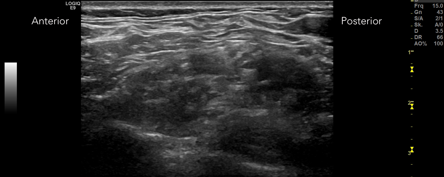
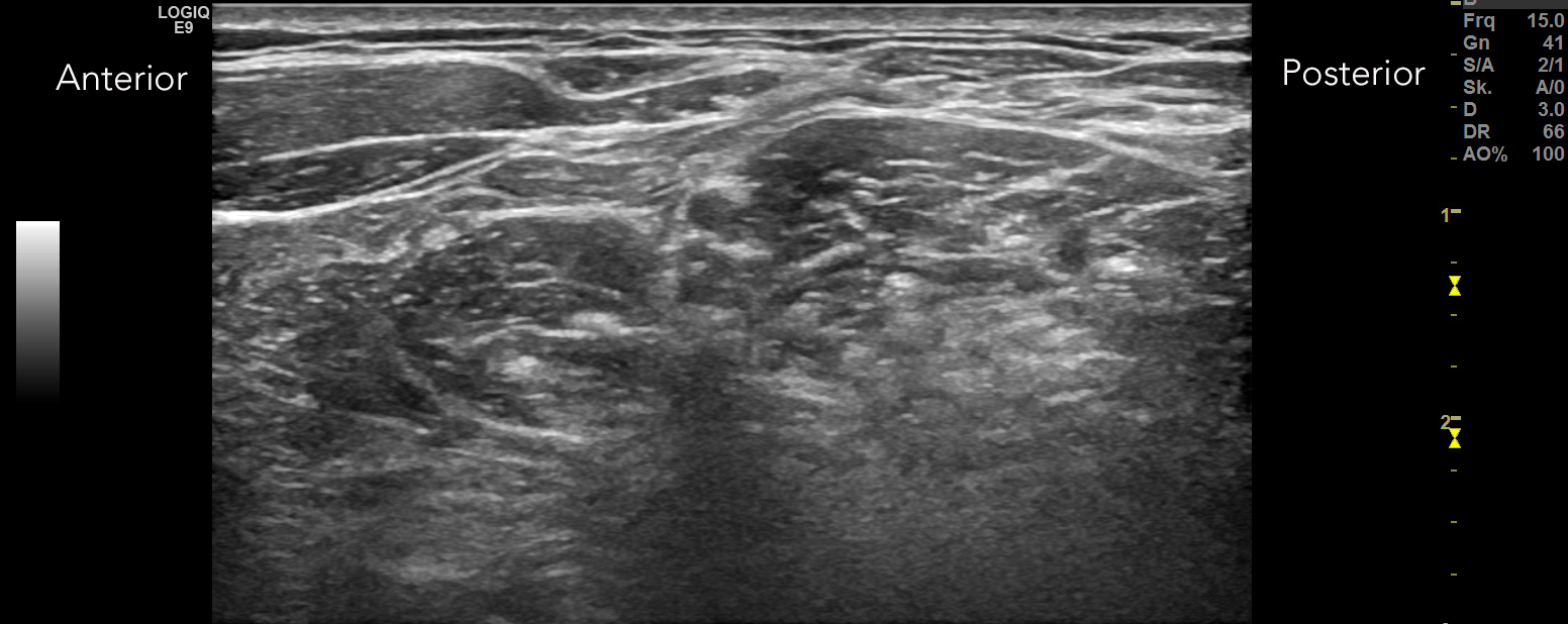
You decide to undertake a ultrasound of the supraclavicular brachial plexus and see the following
(NOTE: the patient is young and slender – you expect optimal requirements for scanning):
Right brachial plexus – interscalene view:

Left brachial plexus – transverse interscalene view:

Left brachial plexus – transverse supraclavicular view:


Do you notice anything? Does it support your suspected diagnosis?
For easier interpretation of the images we built the images below. They show:
a. the image obtained in the patient together with a graph
b. an image obtained in a healthy control
Right brachial plexus – transverse interscalene view:

Left brachial plexus – transverse supraclavicular view:

This case is a beautiful example of how ultrasound can aid the diagnosis in early GBS. You can easily SEE the inflammation of the cervical roots and the trunci, their washed out borders, which make it hard to differentiate the singular fascicles and also differentiation from the surrounding tissue.
Do you have any own experience? We are happy to hear about it!


Recent Comments